

CASE OF THE WEEK – “Fibrous Dysplasia in Maxillary Bone: Case Report” Dr Piyush Somani HOD & Specialist, Gastroenterology, NMC Royal Hospital Sharjah
Gastroesophageal junction Duplication cyst diagnosed by Endoscopic Ultrasound (EUS)
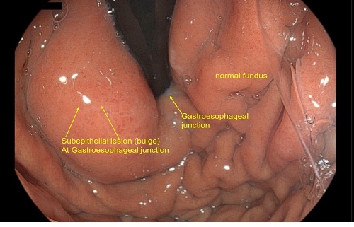
38 years old male was referred for endoscopic ultrasound for subepithelial lesion (bulge) detected at Gastro-esophageal junction on upper gastrointestinal endoscopy.
Upper gastrointestinal endoscopy revealed one large, oval shaped subepithelial lesion (around 32 mm X 15 mm) at Gastroesophageal junction extending to the fundus along the greater curvature towards the posterior wall with normal overlying mucosa.
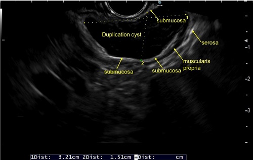
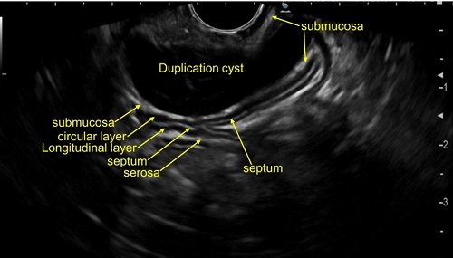
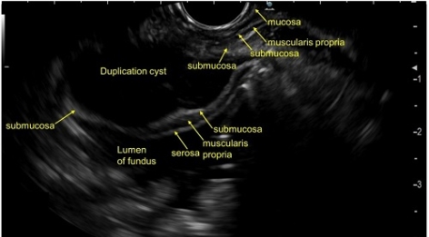
The Endoscopic ultrasound (EUS) revealed anechoic well defined cystic lesion ( 32 mm X 15.1 mm) arising from the submucosa (third layer) at the gastroesophageal junction. The cyst wall consisted of 3 layers ie, muscularis mucosae, submucosa and muscularis propria which favors the diagnosis of duplication cyst. The muscularis propria of the cyst was continuous with the muscularis propria at the gastroesophageal junction. There was no obvious solid component inside the cystic lesion. FNAC/Biopsy was not performed in view of cystic lesion. Patient was reassured about the benign nature of the lesion.
The differential diagnosis of Gastro-esophageal junction subepithelilal lesions include Gastrointestinal stromal tumour, neuroendocrine tumour, leiomyoma, duplication cyst and ectopic pancreas. EUS is the investigation of choice for subepithelial (submucosal) lesions in the entire gastrointestinal tract as CT/MRI will not reveal the origin of the wall layer. The wall layers origin defines the nature of lesion. There are five layers of the wall in the gastrointestinal tract 1) mucosa 2) muscularis mucosae 3) submucosa 4) muscularis propria and 5) serosa. Leiomyoma arises from the 2nd and 4th layer. Neuroendocrine tumour, duplication cyst and ectopic pancreas arises from the 3rd layer. Gastrointestinal stromal tumour arises from the fourth layer.
EUS is a minimally invasive day-care procedure to make diagnosis in subepithelial (submucosal) lesions.
EUS is the diagnostic investigation of choice to investigate duplication cysts since it can distinguish between solid and cystic lesions. EUS shows duplication cysts as anechoic, homogenous lesions with regular margins arising from the submucosal layer or extrinsic to the gut wall. On EUS, duplication cyst wall usually consist of three–five layers and the internal contents may be anechoic or hypoechoic. EUS fine needle aspiration (FNA) is usually reserved for lesions of indeterminate appearance, suspicious for malignancy, or atypical in appearance for duplication cysts.
Reference:
Somani P, Sharma M. Endoscopic ultrasound of esophageal duplication cyst. Indian J Gastroenterol. 2016 Nov;35(6):497-498.